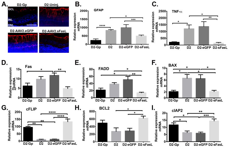
Fig 7. Muller glial cell activation and induction of inflammatory and apoptotic mediators in the retina of D2 mice treated with AAV2.sFasL.
(A) Representative confocal microscopy images of paraffin embedded retinal sections taken from D2-Gp, D2-uninj. D2-AAV2-eGFP, and D2-AAV2-sFasL mice at 10 months of age and stained for GFAP (red) and DAPI (blue) (Scale bar, 100μm). Quantitative RT- PCR was performed on the neural retina isolated from D2-Gp, D2-uninj., D2-AAV2-eGFP, and D2-AAV2-sFasL mice at 10 months of age to quantitate mRNA levels of (B) GFAP, (C) TNFα, (D) pro-apoptotic mediators Fas, FADD, and BAX, and (E) anti-apoptotic mediators cFLIP, Bcl2, and cIAP2. N=5–6 per group. Error bar indicates SEM. *P<0.05, **<0.01, ***P<0.001, ****<0.0001.

