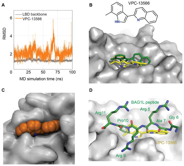
Figure 2. Molecular dynamics simulations of VPC-13566 binding to the AR ligand binding domain.
A- The observed root mean square deviation (RMSD) values for VPC-13566 (orange) and backbone atoms of AR LBD residues (grey) during 100 ns MD simulations. B- Initial docked conformation (green) and representative docking pose of VPC-13566 obtained from MD simulations (yellow). C- Surface representation of BF3 pocket (grey) and VPC-13566 (orange) showing that the ligand fits well into the pocket. D- Superimposition of Bag1L peptide, GARRPR (green) and VPC-13566 (yellow) into the BF3 pocket (grey).

