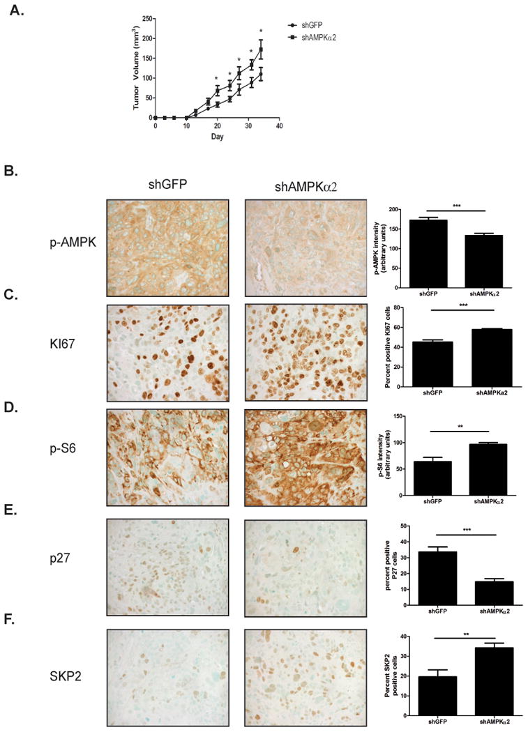
Figure 5. Knockdown of AMPKα2 in HT1376 cells promotes tumor growth in a xenograft model.

A. HT1376 shGFP and HT1376 shAMPKα2 (n=9/group) cells were injected into the flank of NSG mice. Tumor volume was measured twice weekly. B-F. pAMPK(B.), KI67(C.), p-S6(D.), p27(E.) and SKP2(F.) immunohistochemical staining representative images and corresponding quantification (n=9/group). Statistical significance indicated as; *p<0.05, **p<0.01, ***p<0.001
