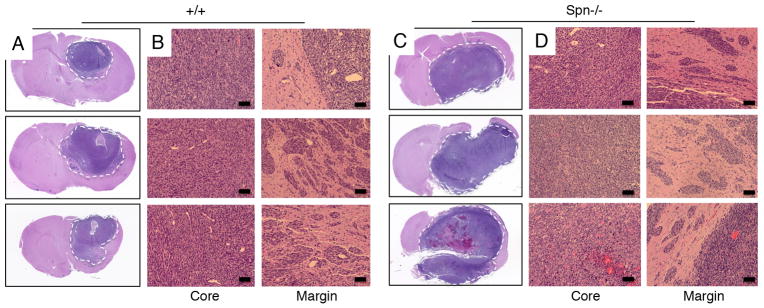
Figure 4. Spn−/− mosaic mouse models display enhanced invasive growth in vivo.
(A–D); Brains of mice harboring tumors generated from wild type (A, B) or Spn−/− (C, D) transformed mouse astrocytes were sliced coronally, embedded in paraffin, and sections were stained with H&E. Note that tumors derived from Spn−/− transformed cells are significantly larger than wild type tumors. Spn−/− cells show more robust invasion away from the primary mass, often reaching the pial surface of the brain (arrows in D).

