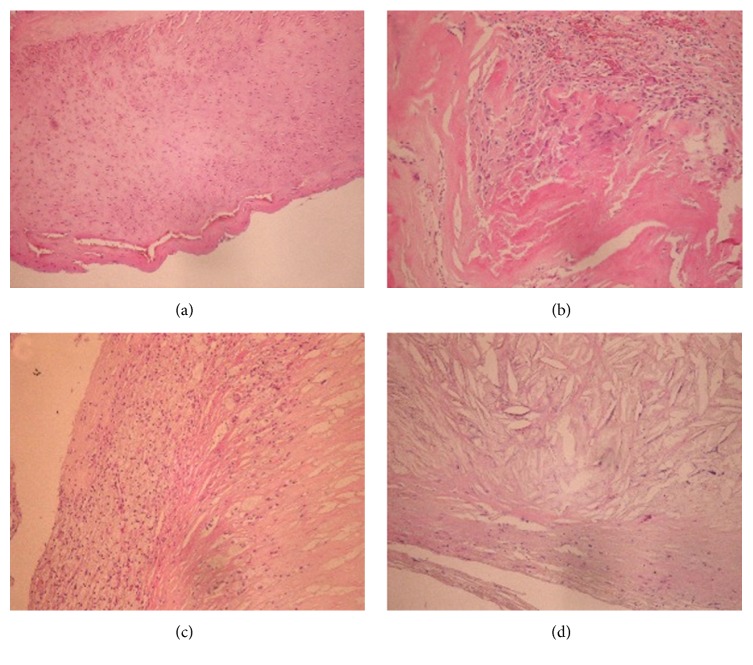
Figure 1.
H&E stained peripheral artery specimens. Samples were submitted to H&E staining, and representative sections are shown. (a) Homogeneous femoral artery and extracellular matrix (magnification ×100). (b) Carotid artery infiltrated by vascular smooth muscle cells and inflammatory cells (×400). (c) Carotid artery showing foam cells (×100). (d) Carotid artery containing cholesterol crystals dispersed in the tissue (×400).

