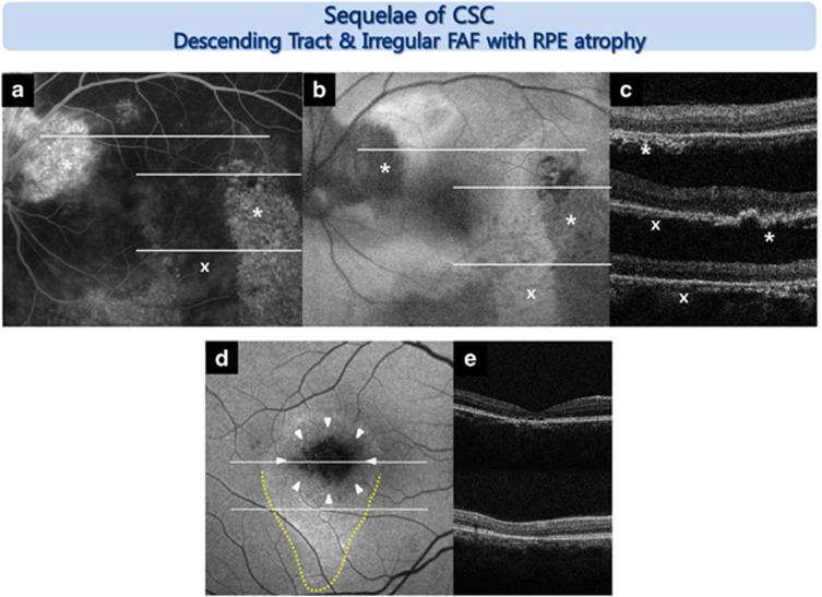
Figure 2.
Representative FAF images from an eye with sequelae of CSC. Descending tract: various FAF changes were observed in the descending tract according to the chronicity of the disease (a–c, d yellow dotted line). The old descending tract (*) showed a window defect in the RPE (area of hyperfluorescence in fluorescein angiography (FA)) and hypo-FAF, corresponding to both RPE and photoreceptor damage as determined by SD-OCT. A recent descending tract (x) showed no window defect in the RPE on the FA image and hyper-FAF, which corresponded to an intact RPE on the SD-OCT image. Irregular FAF with RPE atrophy: focal absolute hypo-FAF and heterogeneous FAF patterns mixing hyper- and hypointensity (d, white arrowhead) over areas of RPE atrophic thinning (e) were observed.

