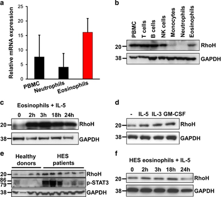
Figure 1.
RhoH expression in peripheral blood eosinophils. Leukocyte subsets were isolated from peripheral blood and RhoH expression was measured by qPCR (a) or immunoblotting (b). Representative immunoblots of eosinophils from healthy donors stimulated with 10 ng/ml IL-5 (c) or 10 ng/ml of IL-5, IL-3 or GM-CSF for 3 h (d), freshly isolated eosinophils from healthy donors or HES patients (e), or IL-5 stimulated eosinophils from HES patients (f), are presented. Values in panel (a) represent means +/− S.D. Data in panels (b–f) are representative for at least three independent experiments

