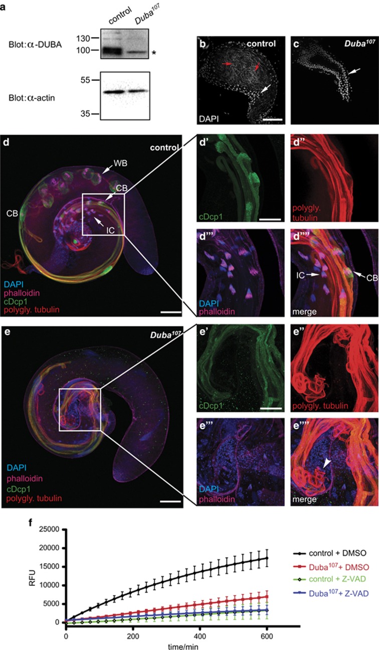
Figure 4.
Testes from Duba-null flies display morphological defects and reduced caspase activity. (a) Testes lysates from 1- to 2-day-old homozygous Duba107flies or controls were immunoblotted to confirm a complete lack of DUBA protein. * indicates a nonspecific signal of the α-DUBA antiserum. Actin levels are shown as loading control. (b and c) Seminal vesicles of Duba107 testes do not contain mature sperm. Seminal vesicles of control (b) and Duba107 (c) testes, stained with DAPI, are shown. White arrows indicate round-shaped nuclei from cells of the sheath of the seminal vesicles; red arrow shows single needle-shaped nuclei of individualised spermatids that are only present in control testes. Scale bar: 50 μm. Repeated three times. (d and e) Confocal projections of testes of 1-day-old control (d) and Duba107 (e) flies reveal structural differences in individualising and elongated spermatids. CB, cystic bulge; IC, individualisation complex; WB, waste bag. Testes were stained with DAPI (blue), phalloidin (magenta) and antibodies against active caspase (cDcp1, green) and polyglycylated tubulin (AXO49, red). Enlarged in (d'–d''''): individualisation complex and cystic bulge. Enlarged in (e'–e''''): Elongated spermatids of Duba107 testes lack IC and CB and show degrading cysts (e'''', white arrowhead). Scale bar: 100 μm in (d and e), 50 μm in (d'–d'''') and (e'–e''''). Repeated four times, four to six testes per condition each time. (f) Caspase activity in lysates from Duba107 testes was measured by DEVDase assay in vitro and compared with control lysate or lysate treated with the caspase inhibitor z-VAD-fmk. Means of three independent experiments and S.E.M. are shown. RFU, relative fluorescence units

