Figure 2.
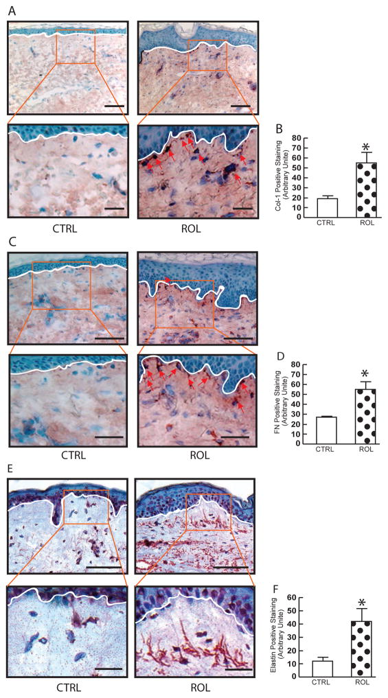
ROL improves dermal ECM microenvironment in aged human skin in vivo. OCT-embedded skin sections (7μm) were obtained from aged (76±6 years) healthy sun-protected buttock skin after topical treatment of vehicle and 0.4% retinol for seven days. (A) Type I procollagen immunostaining. 3× enlargement of the boxed region is shown to lower panels. Representative images of twelve individuals (N=12). Arrows indicate positive cells. Bars=100μm. (B) Quantification of type I procollagen. (C) Fibronectin immunostaining. 2.5× enlargement of the boxed region is shown to lower panels. Representative images of twelve individuals (N=12). Arrows indicate positive cells. Bars= 100μm. (D) Quantification of fibronectin. (E) Tropoelastin immunostaining. 3.0× enlargement of the boxed region is shown to lower panels. Representative images of twelve individuals (N=12). Bars=100μm. (F) Quantification of tropoelastin. All immunostainings were quantified by computerized image analysis (Image-pro Plus software, version 4.1, Media Cybernetics, MD) and data are expressed as mean±SEM, *p<0.05. N=12.

