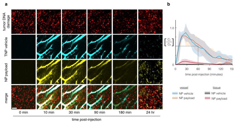
Fig. 5.
Pharmacokinetic analysis of a model nanoparticle shows extended microvasculature half-life and heterogeneous tissue accumulation. Multichannel imaging allows temporal and spatial analysis of both NP and payload distribution. In this example, we monitor the circulation and release of a fluorescent Pt compound from a PEGylated NP [Miller et al., 2015, Nat Commun, 6, 8692]. Nanoparticle concentrations are monitored (a) and quantified (b) using time-lapse confocal fluorescence microscopy in the dorsal window chamber model. Nanoencapsulation extends the initial microvasculature half-life to 55 min, which represents a > 5-fold increase compared to unencapsulated Pt(II) compounds (cisplatin and carboplatin related compounds) in the same animal model [Miller et al., 2014, Chem Med Chem, 9, 1131–5]. Scale bar 50 μm; thick lines and shading denote mean +/− s.e.m. (n=6). Modified from ref. [Miller et al., 2015, Nat Commun, 6, 8692].

