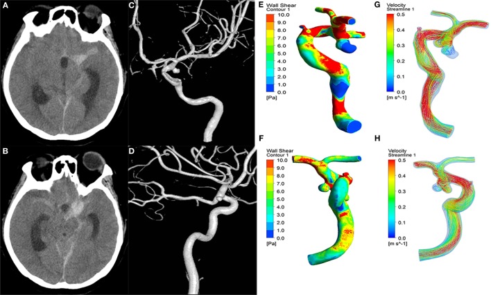
Figure 1.
A 43-year-old female manifested severe headache. First column (A,B): CT showed subarachnoid hemorrhage. Second column (C,D): the 3D-DSA images presented with two aneurysms on the left ICA C7 (ruptured) and right ICA C7 (unruptured). Third column (E,F): wall shear stress (WSS) distribution. The ruptured aneurysms (E) had significantly lower WSS than the unruptured aneurysms (F). Fourth column (G,H): velocity streamlines showing the flow pattern of the aneurysms. The flow patterns of the two aneurysms were simple at peak systole.

