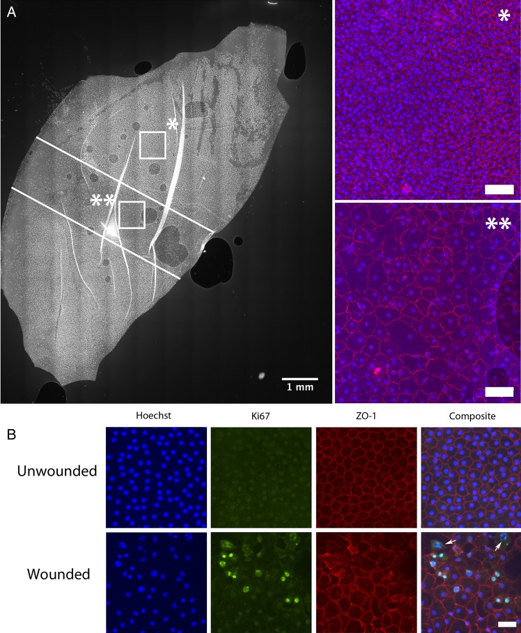
Figure 2.
(A) Photomicrograph showing a Descemet's membrane endothelial keratoplasty (DMEK) graft from half a cornea. A standard injury is created with a silicon-tipped cannula (white lines). After 4 days in standard organ culture media, the wound has been completely covered by migrating endothelial cells. Cell density in the unwounded (*) area is much higher than in the wounded area (**). Tight junctions are not fully formed at 4 days in the wound area (ZO-1 in red). Scale bar 100 µm. (B) Half DMEK grafts were stained for makers of proliferation. Upper panel: Endothelial cells in unwounded areas of the half DMEK graft preserve a healthy, mature monolayer, confirmed by a normal staining for ZO-1. No Ki-67-positive (proliferating) cells are seen. Lower panel: Image taken from the wound site. Ki-67-positive nuclei (green) and cells in various stages of mitosis (white arrows) are seen. In spite of proliferation, cell density remains lower than in the unwounded areas. Scale bar 50 µm.

