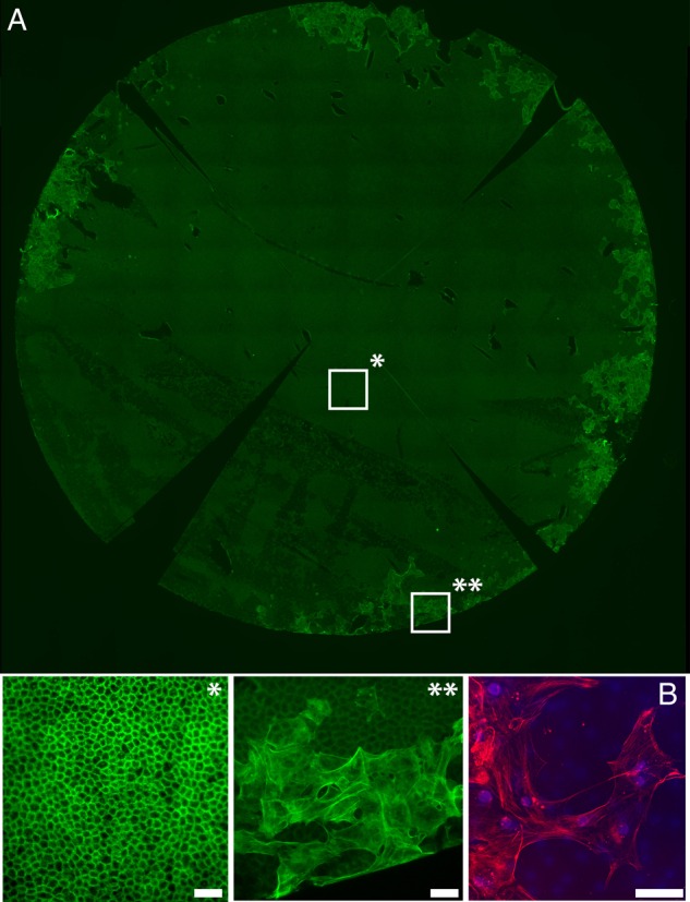
Figure 3.

(A) Fluorescence image showing whole Descemet's membrane endothelial keratoplasty (DMEK) graft that has been peeled, laid back on to the stroma and stored for a further 4 days in standard organ culture media. The graft had been stained with phalloidin (green). *Enlarged segment from a central portion of the graft showing a healthy, hexagonal endothelial monolayer, with a normal actin staining pattern. **Enlarged segment from the periphery of the graft is shown. Large endothelial cells have migrated onto the stromal surface of the DM, with an out-of-focus layer of normal endothelial cells seen underlying these cells on the correct side of the DM. Cells have enlarged, flattened and acquired multiple linear stress fibres, indicating endothelial-to-mesenchymal transformation. (B) A confocal image of the migrating cells shows positive staining for α-SMA, a marker for endothelial-to-mesenchymal (red). Scale bars 50 µm.
