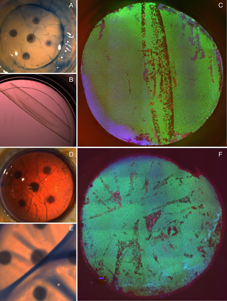
Figure 4.
(A) A branching pattern of cell death, staining positively with trypan blue and corresponding to tissue folds, is seen immediately after preparation. (B) Scrolled graft is shown laying on a glass cover slip in a 48-well culture dish. (C) Global endothelial viability assessment showing characteristic lines of cell death corresponding to the long axis of the scroll. (D) Trypan blue staining immediately after bubble separation shows patterns of cell death consistent with tissue folds. (E) Trypan blue staining after tissue storage shows death cells at the peaks of tissue folds. (F) Global endothelial evaluation of bubble separated graft after trephination.

