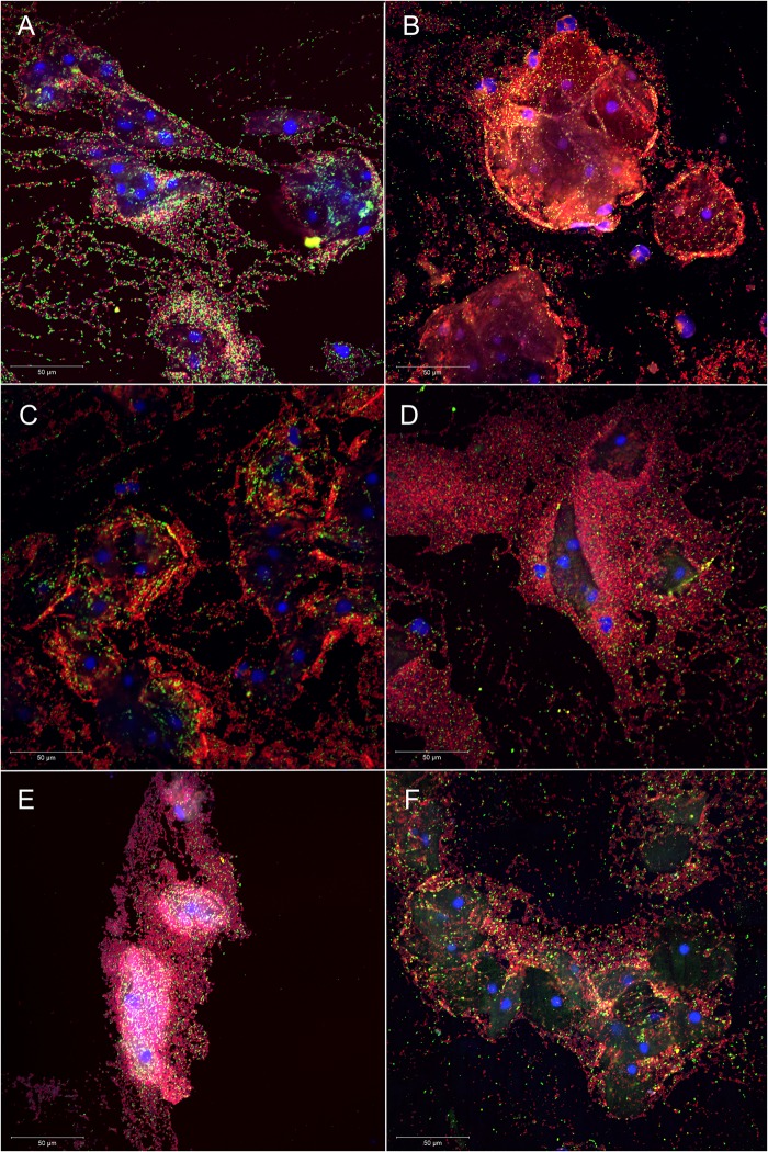
Figure 1.
Superimposed confocal laser scanning images with 400× magnification of Atopobium vaginae+Gardnerella vaginalis biofilm in six vaginal samples (A–F): vaginal epithelial cells DAPI in blue, A. vaginae-specific peptide nucleic acid (PNA)-probe AtoITM1 with Alexa Fluor 488 in green and G. vaginalis-specific PNA-probe Gard162 with Alexa Fluor 647 in red. For clarity, we omitted the BacUni-1 plane, such that the bacteria that did not hybridise with Gard162 and AtoITM1 are visible in DAPI blue only.

