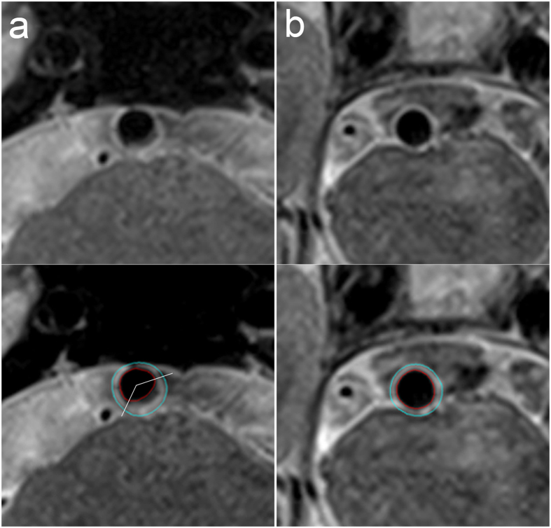
Figure 2. Magnetic resonance images of the vessel wall of a basilar artery plaque.
The lumen area and wall area as measured using the MRI-PlaqueView software are 15.03 mm2 and 24.21 mm2 for the lesion slice (a) and 16.04 mm2 and 13.85 mm2 for the reference slice (b). The maximum wall thickness and minimum wall thickness measured using MRI-PlaqueView for the lesion slice are 2.24 mm and 0.75 mm. The distribution range of the plaque measured using RadiAnt DICOM Viewer software is 133.8°. Therefore, the remodeling ratio, eccentricity index, and plaque range for this plaque are 1.31, 0.67, and 133.8°, respectively.

