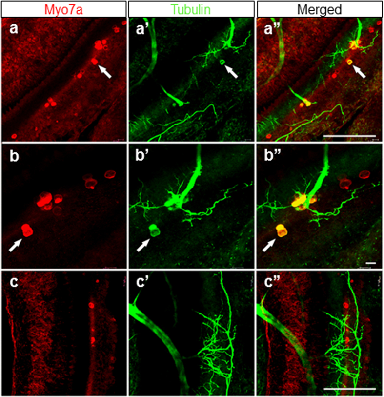
Figure 6. Some HCs are positive for both Myo7a and tubulin.
These single or groups of Myo7a positive HCs of E18.5 Sox2 CKO mice show a patchy distribution (a,b,c) and an unusual pattern of innervation (a’,b’,c’). Note that most fibers are targeted toward Myo7a positive HCs, others are sometimes widely distributed in the topological equivalent of the organ of Corti. (a”,b”,c”) Some Myo7a positive cells are also positive for antibody directed against tubulin, normally a reliable neuronal marker in the ear. Scale bars: 100 μm, except b,b” that indicates 10 μm.

