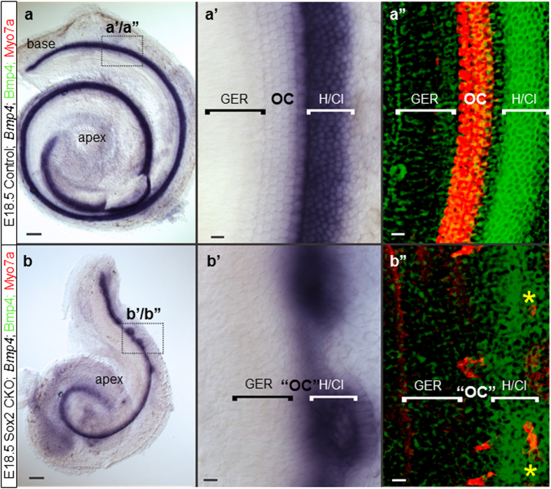
Figure 7. The cellular boundaries of the inner ear are changed in the Sox2 CKO.
(a,a’,b,b’) Loss of Sox2 results in aberration of Bmp4 expression. Instead of being separated by the organ of Corti from the GER, Bmp4 expression is adjacent to the GER. (b, b’,b”) Only the base shows rings of lateral Bmp4 expression and Myo7a positive HCs are both in the center of these rings as well as at the boundary between GER and Bmp4 domain (b”; yellow asterisks). (a’-a”) In controls, the Bmp4 expression in Hensen/Claudius cells is always lateral to the organ of Corti. Scale bars: 10 μm except 100 μm in a and b. GER, greater epithelial ridge; H/Cl; Hensen/Claudius cells; OC, organ of Corti; “OC”, atypical organ of Corti in the mutant.

