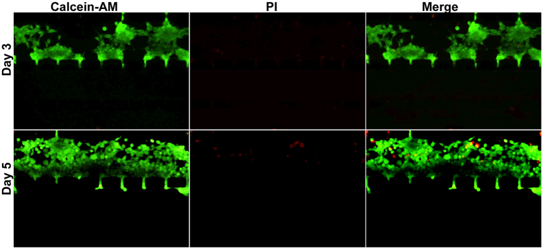
Figure 3. Cell viability assay within the device.
Human embryonic kidney HEK-293 cells were seeded and grown in the cell culture channel of the Matrigel-loaded device under the 2–10% FBS gradient with a flow rate of 3 μL/h. After 3 days (upper) and 5 days (lower) of culture, the cells were subjected to live/dead staining. Living cells were stained green by calcein-AM, and dead cells were stained red by PI (original magnification ×100).

