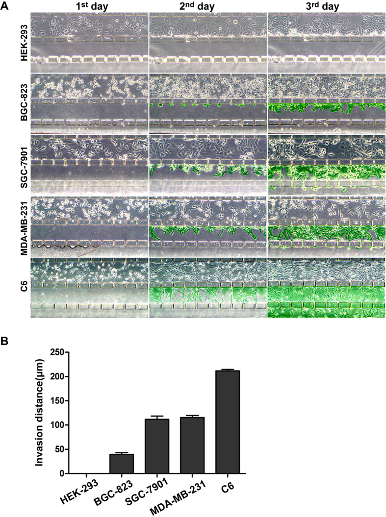
Figure 4. Evaluation of the ability of the microfluidic system to resolve cell invasiveness.
Five different types of cells (C6, MDA-MB-231, SGC-7901, BGC-823 and HEK-293) were cultured in the Matrigel-loaded device under a 2~10% FBS gradient with a flow rate of 3 μL/h. (A) Images of cell invasion were taken every day by phase-contrast microscopy. The cells invading out of the culture channel are indicated by green. (B) The invasiveness of different cell lines was evaluated quantitatively according to the invasion distance of the leading cells (original magnification ×100). Each independent experiment was repeated three times.

