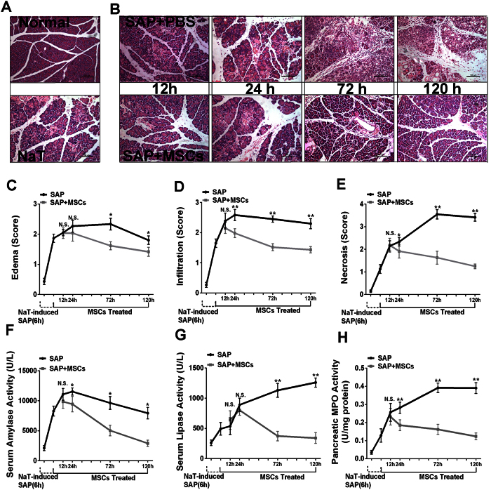
Figure 1. Effects of hMSCs on NaT-induced alterations of pancreatic pathology and time course of cell markers.
(A) Representative hematoxylin/eosin (H&E) stained sections of pancreas from a control mouse demonstrate that the histological features of the pancreas showed typical normal architecture. In contrast, pancreas sections of NaT-treated mice exhibit tissue injury characterized by pancreatic edema, extravascular infiltration and acinar cell necrosis. (B) Pancreas sections from SAP mice at 12, 24, 72 and 120 h after receiving 2 × 10⁶ hMSCs show fewer histological alterations. Original magnification: ×200. (C,D,E) Histological analysis of pancreatitis severity. (F,G,H) Activities of amylase (U/L), lipase (U/L), and MPO (U/mg). Each value represents the mean ± standard deviation (n = 4 or 6 per group). N.S., not significant, *P < 0.05 and **P < 0.01, in comparisons between SAP control groups and SAP + hMSC groups at different time points.

