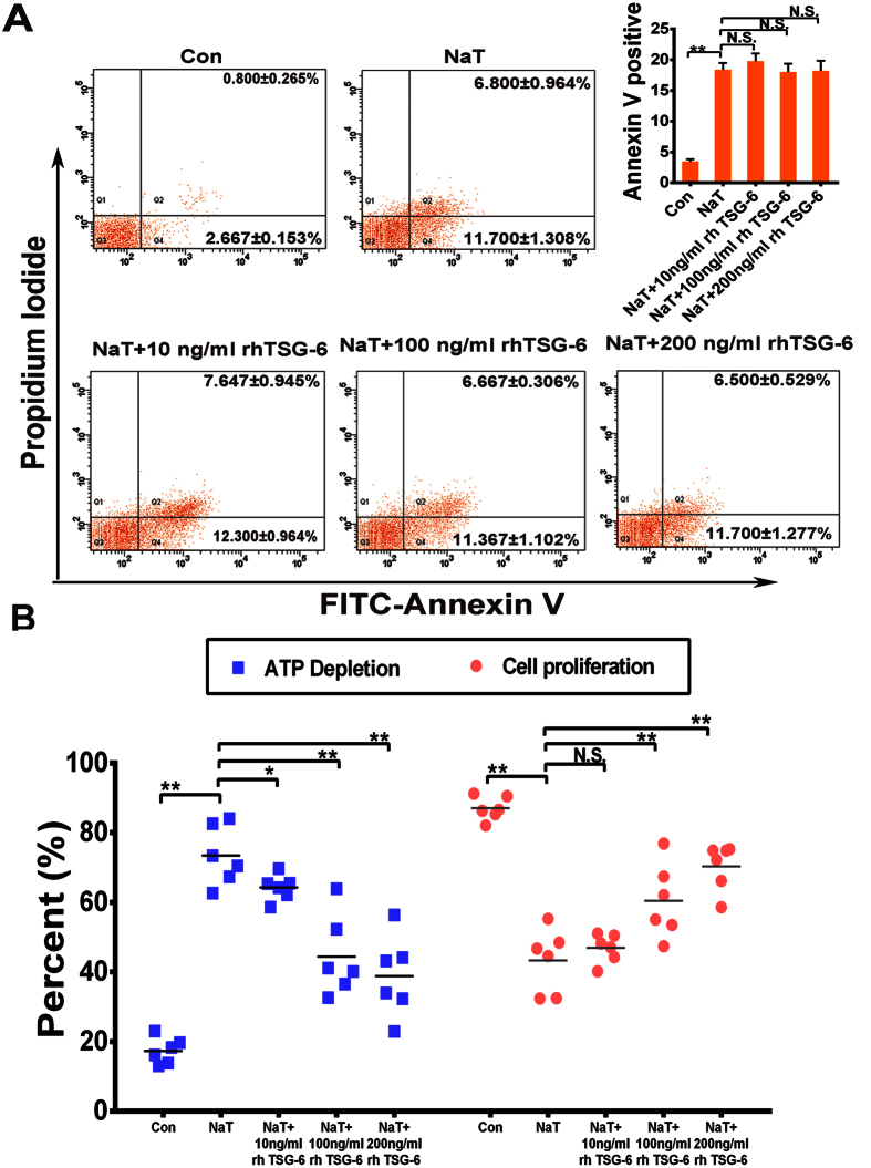
Figure 5. rhTSG-6 improved the activity of NaT-treated pancreatic acinar cells.
(A) In vitro, acinar cells in different group were collected and stained with annexin V-FITC/propidiumiodide and detected by flow cytometry. The right panel indicates the apoptotic cells. Figures are representative of three independent experiments. Each value represents the mean ± standard deviation. N.S., not significant and **P < 0.01, compared with NaT-treated groups. (B) Acinar cell necrosis was reflected by analysis of ATP levels. rhTSG-6 significantly decreased the depletion of ATP in injured acinar cells in a dose-dependent manner, (C) and increased proliferation in a dose-dependent manner. Each value represents the mean ± standard deviation (n = 6 per group). N.S., not significant, *P < 0.05 and **P < 0.01, compared with NaT-treated groups.

