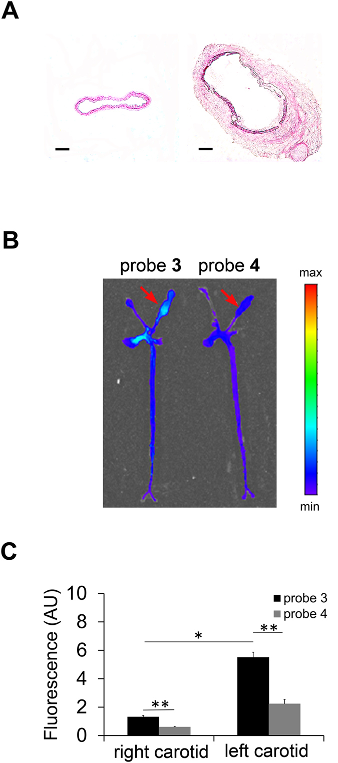
Figure 6. MMP-12 imaging of carotid aneurysm in apoE−/− mice.

(A) Representative H&E staining of NaCl-exposed right (left image) and CaCl2-exposed left (right image) carotid arteries in apoE−/− mice at 4 weeks after surgery to induce aneurysm (scale bar 100 μm). (B,C) Representative fluorescent images (B) and quantitative analysis of fluorescent signal (C) from aortae and carotid arteries harvested at 1 h after intravenous administration of probe 3 (n = 3) or 4 (n = 4). *p < 0.01, **p < 0.001. Arrows point to aneurysmal left carotid artery. AU: arbitrary units.
