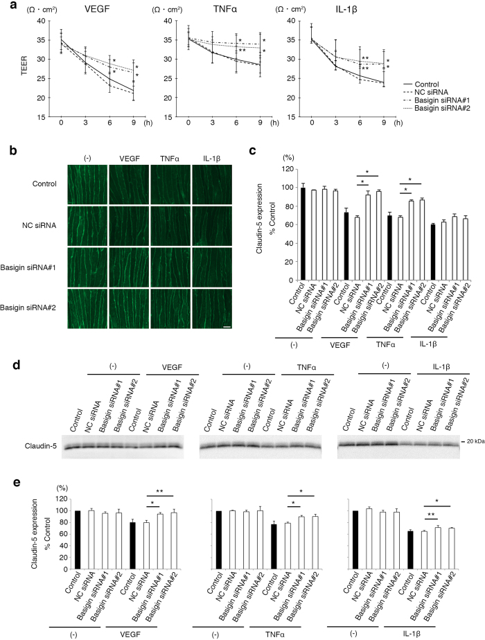
Figure 1. Involvement of basigin in inflammation-induced impairment of neural vascular barrier.
(a) TEERs of bEND.3 monolayers under the stimuli of VEGF, TNFα or IL-1β. Decrease in TEERs by VEGF, TNFα or IL-1β is suppressed in cells transfected with basigin siRNAs, and the differences become significant 6 hours after stimulation. (b,c) Immunofluorescent images of claudin-5 (b) and their corresponding quantitative analyses for cell membrane-localized claudin-5 (c) in bEND.3 cells after the treatment with VEGF, TNFα, or IL-1β for 6 hours without or with the suppression of basigin expression by specific siRNAs. Differences in immunofluorescent intensities for membrane-localized claudin-5 between the cells with NC siRNA and with basigin siRNAs are statistically significant under the stimulation with either VEGF or TNFα, while not significant under IL-1β stimulation. (d,e) Western blot analyses (d) and their corresponding quantitative analyses (e) for cell membrane-localized claudin-5 isolated from bEND.3 cells through in situ biotinylation of cell surface molecules. Cells under the stimuli of either VEGF, TNFα or IL-1β for 6 hours are rescued from the decrease in amounts of cell membrane-localized claudin-5 by basigin siRNAs. Error bars indicate s.d. *P < 0.01; **P < 0.05; NC siRNA, non-silencing siRNA for negative control; Scale bar in (b), 10 μm.

