
Fig. 3.
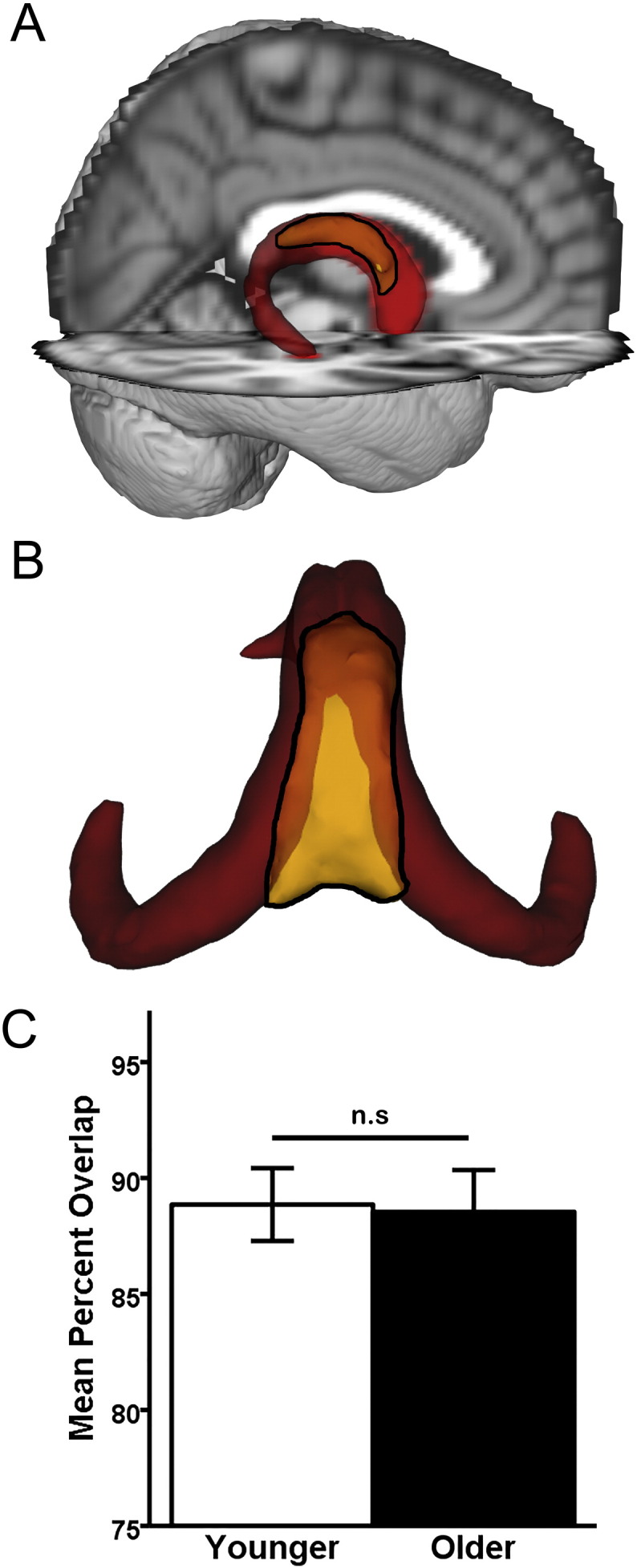
Anatomic validation results. A–B: Qualitative results. The younger-older template (red) and the manually-traced (MT) fornix (yellow with black outline). Opaque yellow within the black outline indicates overlap with the merged younger-older template, while brighter yellow within the black outline are unique to the MT fornix body. A: Visual comparison reveals extensive overlap between the merged younger-older template and the MT fornix body, overlaid on the MNI152 T1 1 mm brain for anatomic reference. B: Caudo-dorsal view of the merged younger-older template and MT fornix body reveals that the main area unique to the MT fornix is in the dorsomedial section (bright yellow), which primarily consists of hippocampal commissural fibers running perpendicular to the fornix body/crus. The more lateral and rostral aspect of the fornix body (opaque yellow) overlap with the merged fornix template. C: Quantitative results. An independent samples t-test revealed that the merged younger-older template provides identical coverage in younger and older adults (p = 0.89). Error bars are ± 1 S.E.M. n.s.: p > 0.05.