Table I.
Glycan composition
| Sample | Glycan structure | Relative amount | Illustrationa |
|---|---|---|---|
| Tn-PSM | GalNAcα1-Ser/Thr | 100 |
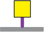
|
| Tri-PSMb | GalNAcα1-Ser/Thr | 28 |
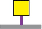
|
| Galβ1-3GalNAcα1-Ser/Thr | 26 |
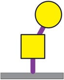
|
|
| Fucα1-2Galβ1-3GalNAcα1-Ser/Thr | 46 |
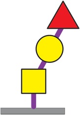
|
|
| STn-OSM | NeuNAcα2-6GalNAcα1-Ser/Thr | 100 |
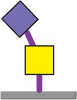
|
| Tn-MUC1 | GalNAcα1-Ser/Thr | 100 |
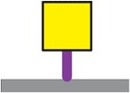
|
| STn-MUC1 | NeuNAcα2-6GalNAcα1-Ser/Thr | 100 |
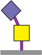
|
| T-MUC1 | Galβ1-3GalNAcα1-Ser/Thr | 100 |
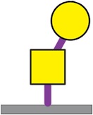
|
| ST-MUC1 | NeuNAcα2-3Galβ1-3GalNAcα1-Ser/Thr | 100 |
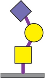
|
aSymbols used: 
b~35–50% of these glycan structures will have the Neu5Gc residue attached to the C6 position of the peptide-linked GalNAc residue (Gerken and Jentoft 1987).
