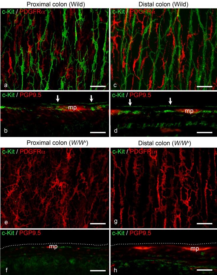
Fig. 1.
a Whole-mount stretch preparation of the subserosal layer stained with anti-c-Kit (green) and anti-PDGFRα (red) antibodies in the wild-type proximal colon. Multipolar ICC (green) and fibroblasts (red) are distributed in the subserosal layer. b Longitudinal cryostat section of the wild-type proximal colon stained with anti-c-Kit (green) and anti-PGP9.5 (red) antibodies. ICC are present in the subserosal layer (arrows) and around the myenteric plexus (mp). c Whole-mount stretch preparation of the subserosal layer in the wild-type distal colon stained with the same antibodies as in (a). Similar findings are observed relative to the proximal colon. d Longitudinal section of the wild-type distal colon stained with the same antibodies as in (b). Similar findings are observed relative to the proximal colon. mp: myenteric plexus; arrows: ICC-SS. e Whole-mount stretch preparation of the subserosal layer in the W/Wv mouse proximal colon stained with the same antibodies as in (a). There are no c-Kit-positive ICC, but multipolar fibroblasts (red) are present in the subserosal layer. f Longitudinal section of the W/Wv mouse proximal colon stained with the same antibodies as in (b). ICC around the myenteric plexus (mp) can be observed, similar to the wild-type colon. There are no c-Kit-positive cells along the subserosal layer (dotted line). g Whole-mount stretch preparation of the subserosal layer in the W/Wv mouse distal colon stained with the same antibodies as in (a). Similar findings are observed relative to the proximal colon of W/Wv mice. h Longitudinal section of the W/Wv mouse distal colon stained with the same antibodies as in (b). No c-Kit-positive cells are observed in either the subserosa or myenteric plexus (mp). dotted line: subserosal layer. Scale bar: 50 µm.

