Figure 9.
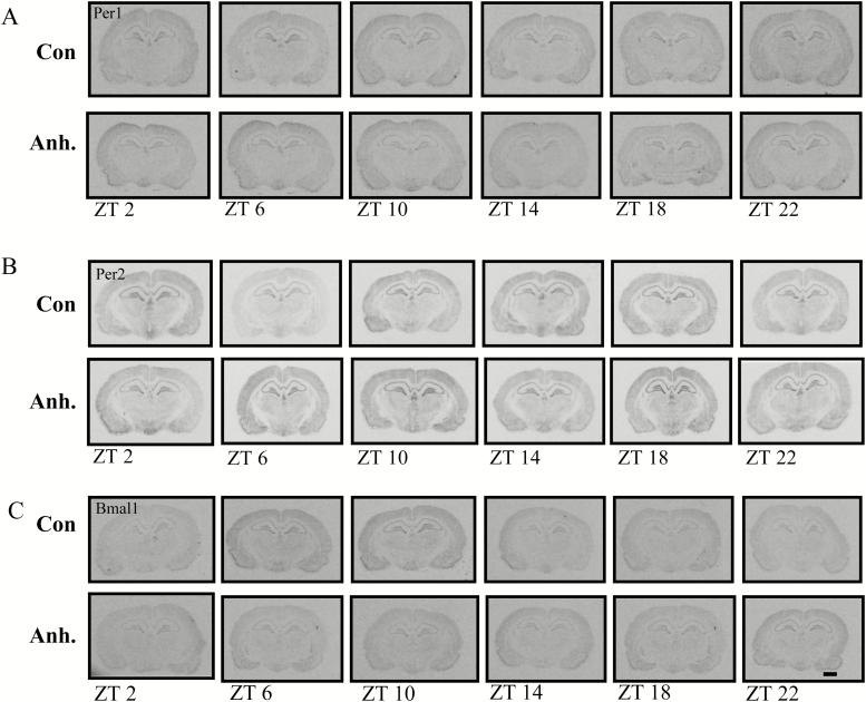
Representative images from the in situ hybridisation histochemistry for analysis of diurnal rhythm of Per1, Per2, and Bmal1 clock genes in coronal sections of the hippocampus in the rat brain. The experimental animals were decapitated after 3.5 weeks of chronic mild stress every 4h. The sampling time is indicated as zeitgeber time (ZT), where ZT 0 is defined as the time point when light switches on. Decapitation was initiated at ZT 22. All animals were housed under a 12h light/dark cycle. Scale bar: applies to all images, 1mm.
X-ray images of the (A) Per1, (B) Per2, and (C) Bmal1 clock genes in the hippocampus in the control groups and the anhedonic-like groups at six time points.

