
Figure 2. Murine plasma MP visualized by Scanning Electron Microscopy.
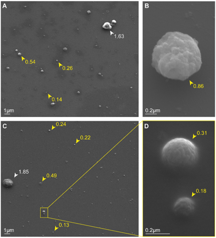
Plasma MP purified from a non-infected DBA/1 mouse have been imaged with a Zeiss Ultra FESEM. (A,C) Magnification x4000. The majority of the visualized vesicles have size corresponding to MP (0.1–1 μm - yellow arrowheads), while only one bigger element (white arrowheads) was visualised on each image probably corresponding to small aggregates of MP or microplatelets. (B,D) Visualisation of plasma MP at a higher magnification, ×23920 and 52160, respectively. Numbers beside arrowheads indicate the measured vesicle diameter expressed in μm.