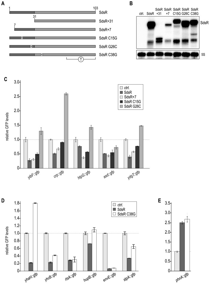
Figure 4.
SdsR employs different binding sites to control target gene expression. (A) Schematic representation of SdsR mutants. Asterisks mark locations of single point mutations; T denotes the terminator region. (B) Expression pattern of SdsR variants. RNA prepared from Salmonella ΔsdsR cells carrying either a control plasmid or expressing SdsR; SdsR+31; SdsR+7; SdsR C15G; SdsR G26C or SdsR C38G from the constitutive PL promoter was analyzed by Northern blotting. Detailed descriptions of all plasmids are provided in Supplementary Table S2. (C–E) GFP fluorescence of Salmonella ΔsdsR cells carrying the indicated gfp reporter fusion in combination with either a control plasmid, or a construct expressing a Salmonella SdsR variant was analyzed by flow cytometry. For each GFP-fusion, fluorescence levels in the presence of the control plasmid were set to 1, and relative changes were determined for cells expressing SdsR sRNA variants. (C) Effect of SdsR+7; SdsR C15G; SdsR G26C on target gene fusions. (D) Effect of SdsR C38G on target gene expression. (E) SdsR C38G activates phnA::gfp expression.

