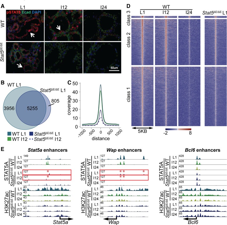
Figure 6.
(A) Decline of active STAT5 in wild-type and mutant mammary tissue. Phospho-STAT5-positive cells in wild-type tissue decreased within 12 h (I12) after terminating lactation and were absent at 24 h (I24). In contrast no pSTAT5-positive cells were observed at I12 in tissue lacking the autoregulatory enhancer Stat5ΔE/ΔE. (B) A total of 5255 out of 9213 STAT5A binding sites (enhancers) in wild-type tissue were shared with Stat5ΔE/ΔE tissue at L1, suggesting that the full establishment of enhancer was not accomplished at lower STAT5 levels. (C) The coverage plot illustrates that the wild-type L1 sample had the highest coverage. Even wild-type tissue at I12 showed a higher coverage than the Stat5ΔE/ΔE samples at L1. Stat5ΔE/ΔE at involution 12 h showed the lowest coverage. (D) Heat map comparing STAT5A coverage in wild-type and Stat5ΔE/ΔE tissue in the three different enhancer categories. (E) Representative examples from the heat map. The STAT5A enhancer in the Stat5a locus was disrupted in Stat5ΔE/ΔE tissue. H3K27ac coverage was reduced but still present. STAT5 binding to the Wap enhancers was greatly reduced in Stat5ΔE/ΔE tissue. Class 3 enhancers were the least affected in mutant tissue. The height of the STAT5A enhancers was lower in the Stat5ΔE/ΔE sample, but H3K27ac remained unaltered.

