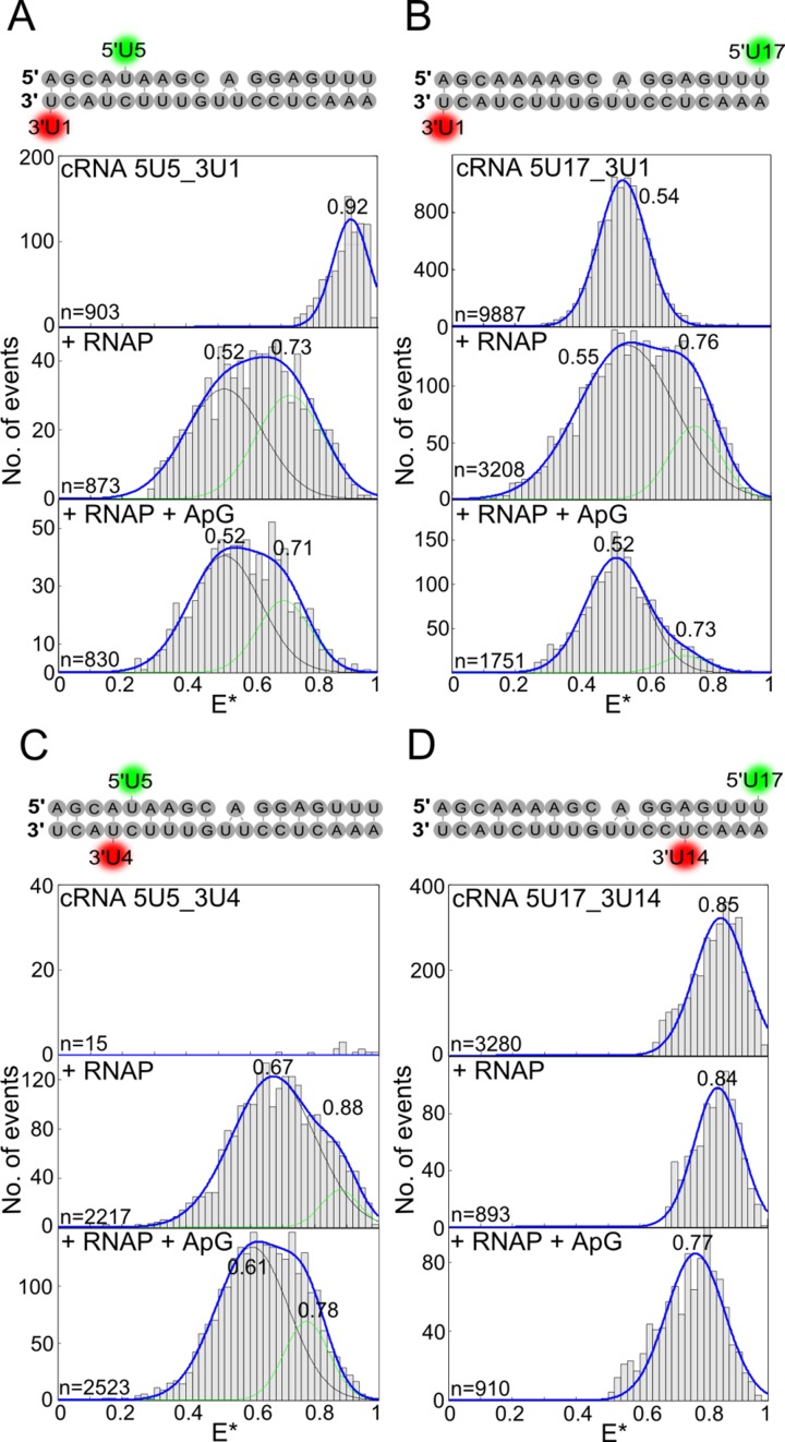
Figure 4.
Influenza virus RNAP-bound cRNA promoter adopts multiple conformations. (A–D) cRNA promoters (sequences and labelling positions depicted in the figure) were labelled with donor and acceptor fluorophores and either analysed alone (top panels), or incubated with a final concentration of 200 nM RNAP (middle panels), or 200 nM RNAP and 500 μM ApG (lower panels) before single-molecule FRET spectroscopy on diffusing molecules was carried out. Histograms from three independent experiments were merged. Ratio E* represents the uncorrected FRET efficiency, n represents the number of molecules and curves were fitted with Gaussian functions to determine the centre of the distributions.

