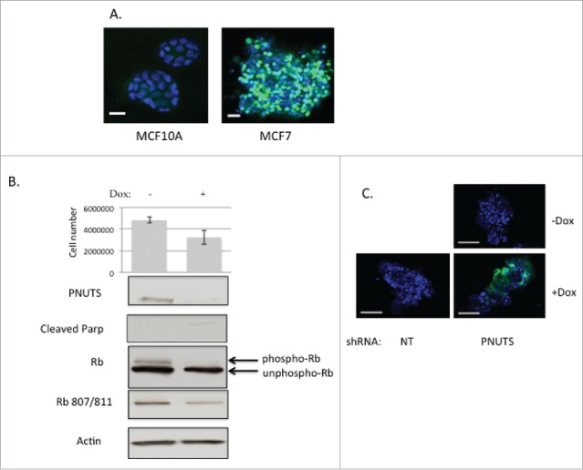
Figure 1.

(A) Non-transformed MCF10A breast epithelial cells (left) and MCF7 breast cancer cells (right) were grown in Matrigel using published methods.34 After 10–12 d in culture, spheroids were analyzed using phosphospecific antibodies to Rb (Ser807/811) according to immunofluorescence protocols (Cell Signaling Technology). Green fluorescence shows phosphorylated Rb. Scale bar, 500 µm. (B) MCF7 cells expressing Dox-inducible PNUTS shRNA were grown in Matrigel cultures for approximately 12 days, +/−Dox stimulation. Cell number experiments were performed by the CellTiter-Glo 3D assay (Promega) with n = 12. Error bars represent standard deviation of the mean and data shown is representative of 5 independent experiments. Cellular protein was obtained from Matrigel cultures by Cell Recovery Solution (Corning). Phospho-Rb and unphospho-Rb are separated by differential migration on SDS-PAGE and are indicated by arrows. Cleaved Parp and Rb phosphorylated at S807/811 is shown. Equal protein loading is shown by Actin immunoblotting. (C) TUNEL assays (Roche) were performed using MCF7 cells grown in Matrigel 3D cell culture expressing NT (nontargeting) or PNUTS shRNA. Dox stimulation is indicated. Data shown is representative of 3 independent experiments.
