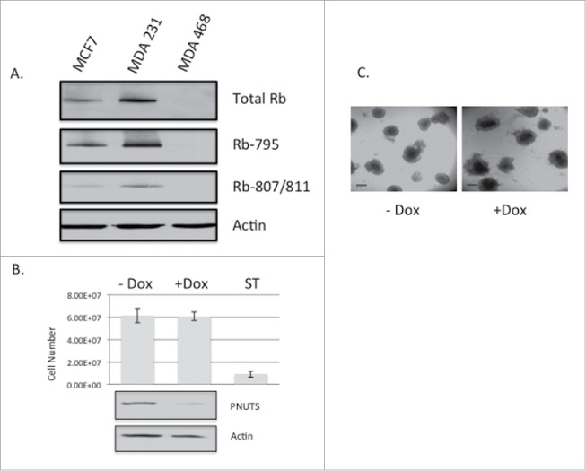
Figure 3.

(A) Immunoblotting with Rb antibodies was performed on lysates from MCF7, MDA-MB-231, and MDA-MB-468 cell lines. Phosphospecific antibodies to Rb on S795 and S807/811 are indicated. Actin immunoblotting is shown as a loading control. (B) MDA-MB-468 cells expressing Dox-inducible PNUTS shRNA were grown in 3D cultures for 12 days, +/− Dox, followed by immunoblotting for PNUTS and Actin. Cell number was determined by the CellTiter-Glo 3D assay (Promega). ST (1mM Staurosporine, 24 hr) was used as a positive control for cell death. Error bars represent standard deviation of the mean of n = 10 and the data shown is representative of 3 independent experiments. (C) Images of MDA-MB-468 cells grown in Matrigel 3D cell culture +/− Dox stimulation show cell morphology. Scale bar, 200µm.
