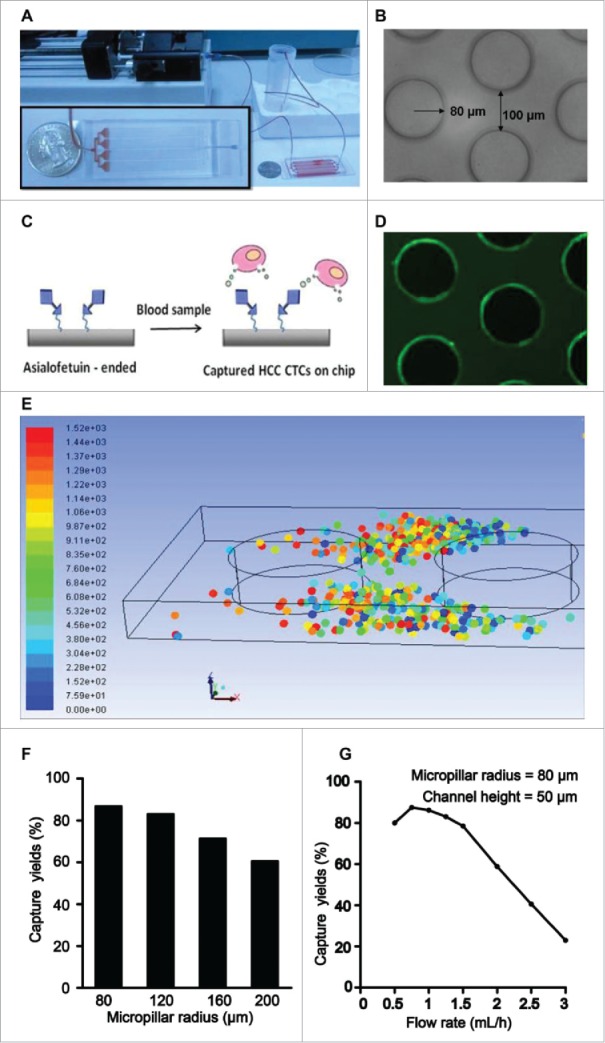
Figure 2.

Representation of the configuration and operational mechanism of an integrated device for capturing circulating tumor cells (CTCs) of hepatocellular carcinoma (HCC). (A) Pictures of the microfluidic system for detection of HCC CTCs. (B) Optical micrograph of the patterned structures inside the chip. (C) Illustration of the HCC CTC detection on chip. (D) Fluoresce micrograph of the structures, which was coated with streptavidin and then introduced with biotin-FITC-IgG. (E) Simulation of the CTC capture inside the microchip. The dots of different color represent captured CTCs. (F) The relation between different micropillar sizes and the capture yieldat a same flow rate of 1 mL/h. (G) The relation between different flow rates and the capture yields with a same micropillar size of 80 µm.
