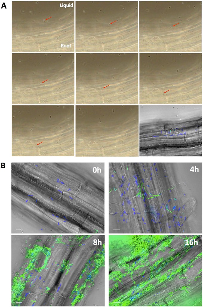
FIG 1 .

(A) Sequential phase-contrast pictures of an A. thaliana root inoculated with B. subtilis NCIB 3610 cells harboring PtapA-yfp and Phag-cfp reporters. The medium used was MSNg, and imaging started immediately after the inoculation. The red arrow points to a cell swimming toward the root and settling on it. Magnification, ×60. Each picture is separated by 0.5 s; the complete movie can be found in Movie S1 in the supplemental material. The last image is a fluorescence picture of the same frame with overlays of fluorescence (false-colored green for YFP and blue for CFP) and transmitted light (gray) images. (B) When B. subtilis cells colonize A. thaliana roots, they first express motility genes, followed by matrix genes. NCIB3610 cells harboring PtapA-yfp and Phag-cfp were coincubated with A. thaliana seedlings and imaged at 0, 4, 8, and 16 h postinoculation. Shown are overlays of fluorescence (false-colored green for YFP and blue for CFP) and transmitted light (gray) images. Pictures are representative of at least 12 independent roots. Bars, 10 μm.
