FIGURE 2.
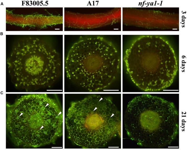
Comparisons of spatiotemporal colonization pattern by A. euteiches in the F83005.5 susceptible line, A17 WT and nf-ya-1-1. For cytological observations of A. euteiches was labeled in green with WGA-FITC (Bar = 100 μm). Infected seedlings were imaged with a GFP long pass filter enabling the oomycete detection in green and the root autofluorescence in red. Root cross sections are 100 μm thick. Oospores are indicated by white arrows. (A) Root surface observation 3 dpi, F83005.5 rhizodermis is fully colonized while a limited amount of hyphae is observed at the surface of A17 and its nf-ya1-1 mutant. A bright red autofluorescence is observed in the last two lines but not in F83005.5. (B) Root sections 6 dpi. A. euteiches hyphae spread intercellularly within the cortex of all three lines. The oomycete colonizes the vascular tissues of F83005.5 as early as 6 days following inoculation whilst these tissues remained free of hyphae in A17 and nf-ya1-1 3 weeks after the inoculation. (C) Root sections 21 dpi. A. euteiches hyphae spread intercellularly within the cortex of all three lines and in the stele of F83005.5. Numerous oospores are being observed in F83005.5 (arrowheads mark some of them) after 21 days, showing the oomycete fulfilled it sexual lifecycle in the susceptible host.

