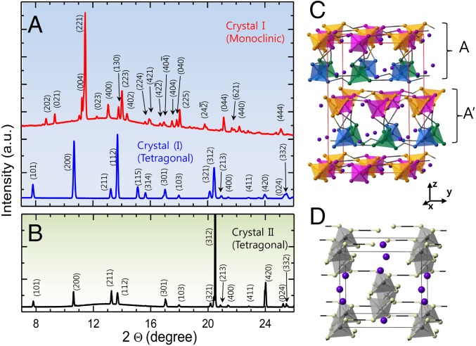
Fig. 6.
In situ X-ray diffraction patterns of KDP crystals formed from supersaturated solutions. (A) Crystal I with monoclinic structure is formed from highly supersaturated solution at S = 4 (Top in A). The monoclinic crystal of crystal I transforms to tetragonal structure later (Bottom in A). (B) Crystal II with tetragonal structure is formed from low supersaturated solution at S = 2.7. X-ray diffraction pattern of the crystal I was refined through a Pawley refinement analysis using Lorentzian profile function. The diffraction patterns for the crystal II coincide exactly with the peak of reported tetragonal phase. Using the obtained structural information and atomic coordinates, the diffraction peaks were indexed. The monoclinic and tetragonal crystal structures are visualized in C and D, respectively (see Crystal Nucleation Analysis for details).

