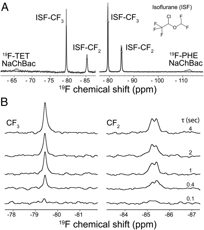
Fig. 3.
19F NMR quantification of isoflurane binding to NaChBac. (A) A set of representative pairwise 19F NMR spectra showing on-resonance (black, Von) and off-resonance (gray, Voff) saturation of the 19F TET-labeled T189C NaChBac (Left) and the 19F PHE-labeled NaChBac (Right). The on- and off-resonance frequencies were –65.6 and –99.0 ppm for the 19F TET (Left), –112.6 and –51.9 ppm for the 19F PHE-labeled NaChBac (Right), respectively. 19F chemical shifts were referenced to the trichlorofluoromethane resonance at 0 ppm. The saturation time was 4 s. (Inset) Isoflurane molecule. (B) Representative 19F STD NMR spectra showing isoflurane interaction with the T189C NaChBac at various saturation times (τ). For each given saturation time, the STD spectrum (VSTD) results from subtracting the on-resonance spectrum from the off-resonance spectrum (VSTD = Voff – Von). (Left and Right) Spectra from the −CF3 and −CF2 moieties of isoflurane, respectively.

