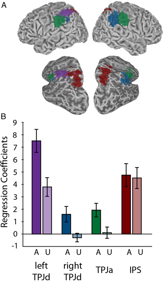
Fig. 3.
Group ICA results. (A) Winner-take-all maps showing location of Left TPJd (purple); Right TPJd (blue); TPJa (green); intraparietal sulcus (IPS) (red). A partially inflated view from a posterior, dorsal, lateral angle is also shown to better reveal the inside of the intraparietal sulcus. (B) Regression coefficients for the aware (A) and unaware (U) conditions. Error bars show SE among subjects.

