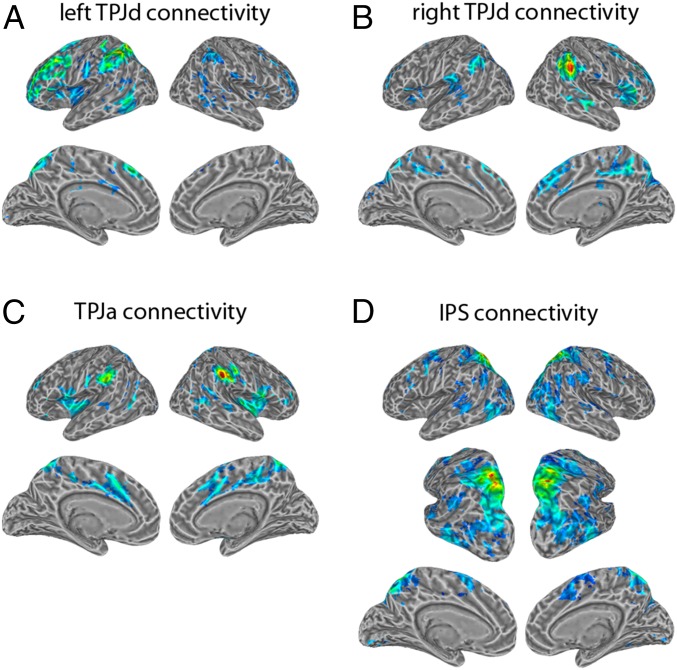
Fig. 5.
Functional connectivity patterns for the independent components obtained in the left TPJd (A), right TPJd (B), TPJa (C), and the intraparietal sulcus (D). The time series for each component served as a seed for its functional connectivity analysis. Shown are regression coefficients (positive only) for voxels that pass a threshold of P < 0.01 corrected for multiple comparisons adjusted for a 5-voxel minimum cluster size. A partially inflated view from a posterior, dorsal, lateral angle is also shown to better reveal the inside of the intraparietal sulcus.

