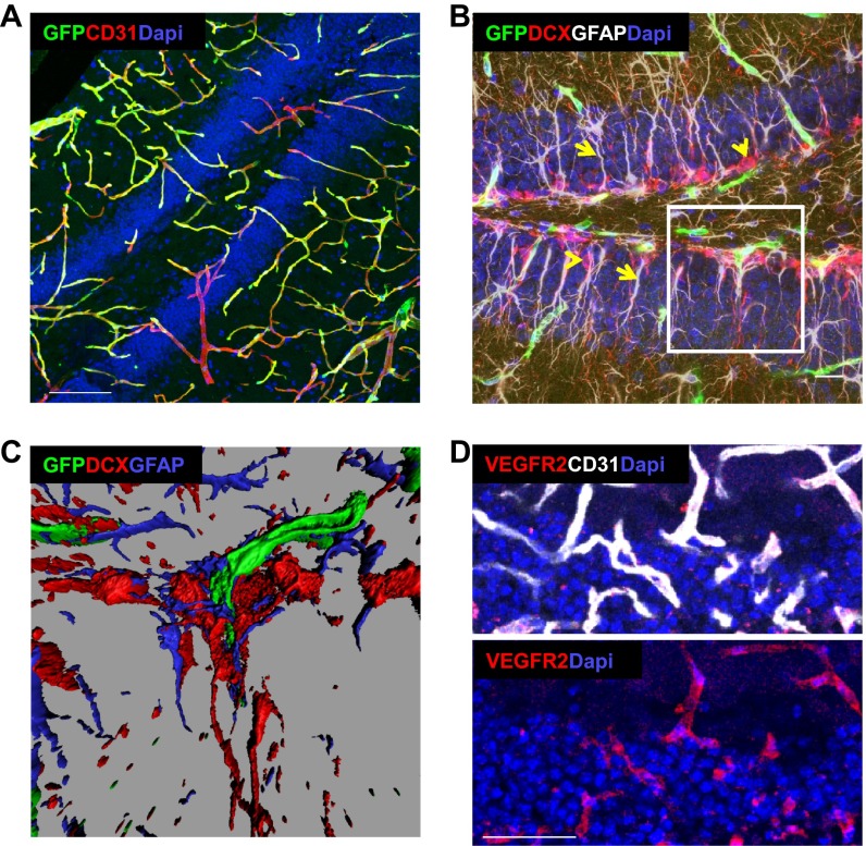
Fig. S4.
VEGFR2 is expressed exclusively in hippocampal ECs. A knock-in VEGFR2-GFP reporter was used to detect VEGFR2-expressing cells. (A) Costaining with CD31. (Scale bar, 100 μm.) (B) Costaining with GFAP, detecting both astrocytes and NSCs (white, arrows) as well as DCX (red, arrowheads). (Scale bar, 20 μm.) (C) Three-dimensional reconstructions of B (Inset) indicate lack of GFP expression in GFAP+ or DCX+ cells, which are found in close physical proximity to GFP+ capillaries. (D) Immunostaining for VEGFR2 in 3-mo-old VEGF mouse induced for the last month. (Scale bar, 50 μm.)

