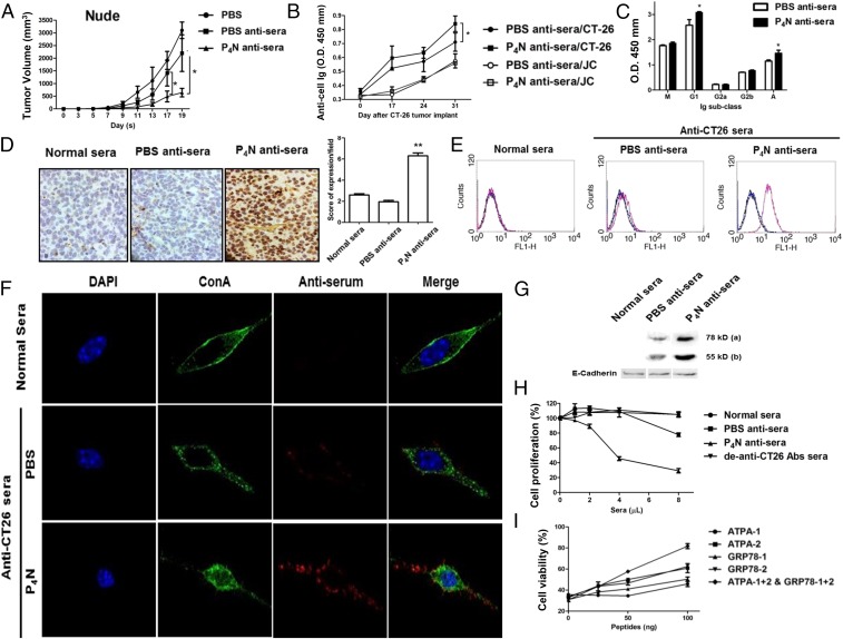
Fig. 3.
Antitumor effect of P4N-induced antiserum in immunodeficient mice and the target antigens of the antiserum. (A) Nude mice bearing CT26 cell tumors (n = 5 per group) were treated with 100 μL of PBS, PBS antisera, or P4N antisera weekly. Tumor volumes were measured every 2 d after treatment. (B) Titers of specific anti-CT26 or JC cell antibodies for the PBS antisera or P4N antisera on days 0, 17, 24, and 31 were measured. (C) Effects of P4N on the changes in the isotypes of specific anti-CT26 cell antibodies were examined. The titers of these antisera (1,600× dilution) were measured for IgM, IgG1, IgG2a, IgG2b, or IgA. (D) IHC analysis of antisera bound to tumor antigens on the surface of CT26. Color development by HRP-conjugated anti-mouse antibodies indicates the antisera antibody. (E) Antisera binding to tumor antigens on the surface of CT26 cells was indirectly detected with FITC-conjugated goat anti-mouse IgG antibody. (F) Subcellular location of antigens recognized by the antisera was monitored by confocal microscopy. Alexa Fluor 568-conjugated anti-mouse Ig antibodies were used to display the presence of antisera (red). Alexa Fluor 488-Con A and DAPI were used to indicate the cell membrane (green) and the cell nucleus (blue), respectively. (G) Normal sera, PBS antisera, or P4N anti-sera were used to probe Western blots of membrane proteins of extracted from CT26 cells. Two bands of 78 kDa and 55 kDa were visualized: E-cadherin proteins were used as a loading control. For all results, significant differences between the P4N antisera group and the PBS antisera group were calculated and labeled with *P < 0.05. (H) Proliferation of CT26 cells treated with 1, 2, 4, or 8 μL of sera was analyzed by MTT assay. To create de–anti-CT26 antibody sera, the anti-CT26 autoantibodies from P4N antisera were presubtracted by CT26 cell binding. The data are reported as the proliferation index. Significant differences between the P4N-treated groups and the untreated group were indicated by *P < 0.05 (n = 5). (I) Cytotoxicity of P4N antisera after neutralization with ATPA and GRP78 peptides. (Magnification: C and F, 400×.)

