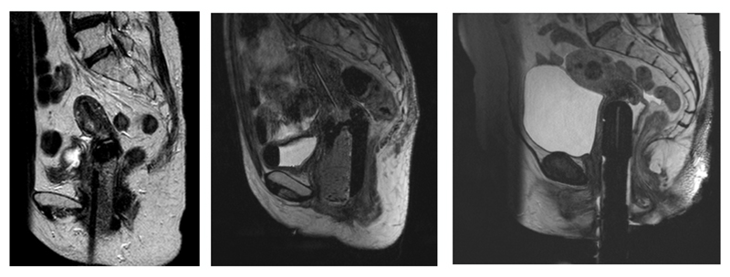
Figure 12.
T2-weighted MR images of three different types of MR compatible brachytherapy applicators, which appear dark in contrast with surrounding tissue. In the central figure, a fluid filled tube appears bright within applicator. In some cases an anterior saturation band is employed to eliminate signals from the abdominal wall, to prevent motion artifacts in the MR images.

