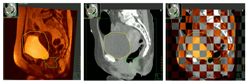
Figure 9.
CT-MR fusion of rectal cancer patient. Both examinations were undertaken using a flat bed and the position of pelvic bones coincides almost perfectly. However, bladder filling is quite different and many soft tissues, including the rectum, are considerably displaced. The benefit of MRI for this particular RT treatment plan is thus limited.

