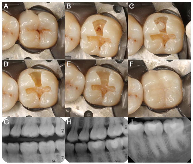
Figure 1. Direct pulp capping on #18 using CH.
(A) Pre-operative clinical photograph of #18. (B) OB preparation with exposed MB pulp horn. (C) Dycal placement on the exposed pulp. (D) Fuji Lining LC placement as a liner directly on the Dycal. (E) Fuji II LC placed as a base on #18. (F) Composite restoration on #18. (G) Pre-operative radiograph of #18. (H) Post-operative radiograph of #18. (I) Periapical radiograph of #18 at follow-up after 2½ years.

