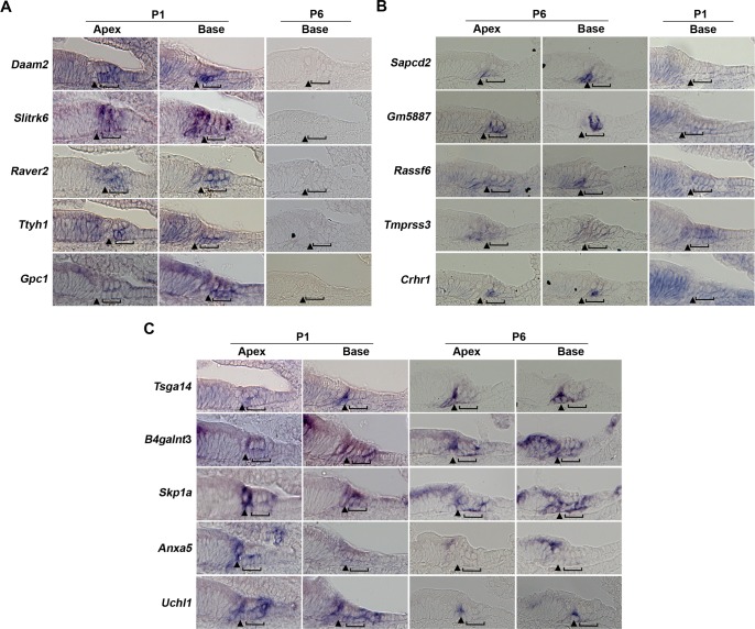
Fig 2. In situ validation of supporting cell-specific transcripts.
Examples of supporting cell genes enriched in (A) P1 supporting cells with basal P6 sections to show negative expression (B) P6 supporting cells with basal P1 sections to show negative expression (C) Both P1 and P6 supporting cells, with sections to show both apical (less mature) and basal (more mature) regions at each age. Brackets: Deiters’ cells; Arrowhead: pillar cell region; horizontal line, greater epithelial ridge region.

