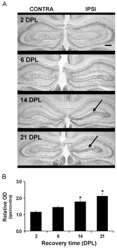
Fig. 2.

Pattern of acetylcholinesterase (AChE) activity histochemical staining of hippocampal cholinergic terminals in response to entorhinal cortex lesioning. (A) Representative photomicrographs of the AChE staining density in the dorsal region of the hippocampal formation, ispilateral (IPSI) and contralateral (CONTRA) to the lesion site at 2, 6, 14 and 21 days post-lesion (DPL). As indicated by the arrows, a significant increase in AChE staining in the outer portion of the molecular layer of the dentate gyrus is observed at 14 and 21 DPL; Coinciding with the replacement of entorhinal cortex projections by septal-hippocampal ones. The black line represents a scale bar of 20 μm in a 2.5 × magnitude photomicrographs. (B) Quantification of AChE staining density corrected for laminar shrinkage; Values are expressed as relative optical density (ipsilateral: contralateral OD) measures of AChE staining with respect to dentate molecular width. Bars correspond to the mean of 5 animals/group±SEM; *p<0.05 as compared to 2 DPL.
