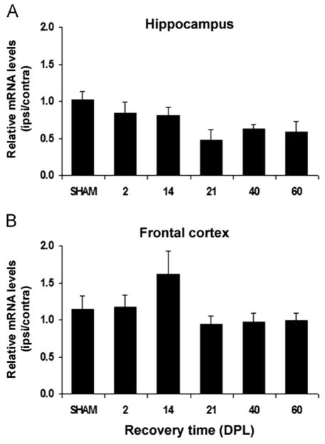
Fig. 4.
Time course of ABCG1 mRNA expression in the hippocampus and frontal cortex of SHAM-operated and lesioned mice following entorhinal cortex lesion (ECL). The ABCG1 mRNA levels normalized to β-actin mRNA levels, are expressed as ipsilateral:contralateral ratios (mean of 5 mice/group±SEM). The relative expression of ABCG1 mRNA in the hippocampus (A) and frontal cortex (B) of lesioned mice are not significantly different from those observed in SHAM-operated mice at any of the observed time points (DPL: days post-lesion).

