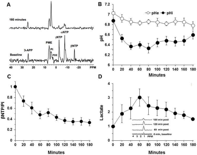
Figure 1. The intracellular pH (pHi), extracellular pH (pHe), bioenergetics and lactate profiles of human melanoma xenograft after lonidamine (LND) administration.
A) In vivo localized (Image Selected In vivo Spectroscopy-ISIS) 31-Phosphorus magnetic resonance spectroscopy (31P MRS) spectra of a human melanoma xenograft grown subcutaneously in nude mice (lower) pre- and (upper) 180 min post administration of LND (100 mg/kg, i.p.). Resonance assignments are as follows, 3-APP (3-aminopropylphosphonate); PME (phosphomonoesters); Pi (inorganic phosphate); PDE (phosphodiesters); γNTP (γ nucleoside-triphosphate), αNTP (α nucleoside-triphosphate), and βNTP (β nucleoside-triphosphate). Decrease in βNTP levels and the corresponding increase in Pi following LND administration (Upper spectrum of panel A) indicating impaired energy metabolism. B) pHi, pHe profiles as a function of time. C) The changes of bioenergetics (βNTP/Pi) (ratio of peak area) relative to baseline D) Change in tumor lactate as a function of time, inset picture showing lactate spectra using 1H MRS with Hadamard Selective Multiquantum coherence transfer pulse sequence in human melanoma xenografts. Area under the curve was compared to baseline at each time point and was normalized to baseline levels as a function of time in response to LND (100 mg/kg; i.p.) administered at time zero. The values are presented as mean ± S.E.M. When not displayed, S.E.M. values were smaller than the symbol size. Part of this research was originally published in NMR Biomed (7) and modified in the current review manuscript.

